The last three decades have seen expeditious advances in types of medical imaging. The advent of diagnostic imaging has allowed radiologists to use high-resolution images of soft and hard tissues to examine health conditions and dictate adequate treatment schemes for patients.
The good news is that, our company operates medical imaging facilities across the country and is the nation’s leading provider of diagnostic imaging services. We provide diagnostic imaging services to health providers and their patients.
Please find out how modern medicine uses different types of medical imaging at its disposal to diagnose diseases and ensure patient safety.
Different Types of Medical Imaging:
Vascular Imaging
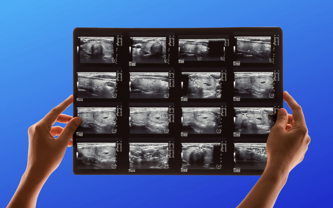
It is a non-invasive, painless, and radiation-free test that detects plaque buildup, aneurysms, and other abnormalities like artery hardening that interfere with blood flow. This technique uses ultrasound technology to provide you and your doctor with an overview of what is happening within your body’s blood vessels.
A high-frequency sound wave travels from the probe into the body via the gel. A sensor collects the sounds reflected from the body.
Photoacoustic Imaging
This imaging technique combines optimal imaging and luminance spectral sensitivities with tissue penetration and ultrasound imaging capabilities. With its unique ability to visualize molecular changes deep within living tissue with an ultrasound-like spatial resolution, photoacoustic imaging may be used to provide a real-time, non-invasive diagnosis of a variety of prevalent diseases.
The robustness of photoacoustic imaging lies in its ability to image molecular and physiological changes at profound levels in living animals.
Thermal Imaging
Physicians use thermal imaging to study disease states like skin inflammation or abnormalities in blood flow associated with clinical abnormalities. Many physicians presently use thermographic imaging to screen for various medical conditions, including muscular pain, arthritis, circulatory problems, and repetitive strain injuries.
3D Medical Imaging
A 3D medical imaging system is a revolutionary optical imaging technique that provides a magnified image of the body’s interior for medical examinations using 3D imaging methods. Radiology, ultrasound, and computed tomography (CT) are part of 3D imaging technology.
In 2020, the global market for 3D medical imaging services was valued at $207,134.9 million, and it is expected to reach $377,062.6 million by 2030, registering a compound annual growth rate of 6.6%.
Molecular imaging (MI)
It helps radiologists quantify, visualize, and describe the subcellular and cellular features of living things.Molecular imaging aims to get detailed pictures of the inside of the body, showing molecular and cellular activities.
Myelogram
Myelograms are used to provide detailed information about your joints that cannot be obtained from MRIs or CT scans. A myelogram usually shows tumors, infections, spine disorders, or problems caused by arthritis.
Arthrogram
An arthrogram is an imaging procedure that directs and evaluates the injection of contrast precisely into a joint using X-rays. An arthrogram identifies joint problems more thoroughly than a conventional medical imaging method, such as a CT scan or an MRI.
Thoracic Imaging
It is a diagnostic imaging technique for chest diseases. According to the American Thoracic Society, the technique is instrumental in imaging COVID pneumonia.
Regardless of the condition that you may suffer from, your physicians can use an imaging tool to diagnose it and start treating you. Contact us about medical imaging.